Cancer Biology: Theoretical Perspectives
Our lab has been involved in the use of genetic screening to uncover genes relevant to cancer etiology and therapy. We look at the cancer problem from the perspective of genetics through which we try to trick cancer cells into revealing their secrets with an eye toward developing new therapies or figuring out how to best used existing therapies. How we view the cancer problem and some of the efforts we have undertaken are described below.
- Recent Studies
- The adaptive immune system is a major driver of selection for tumor suppressor gene inactivation
- Profound tissue specificity in proliferation control underlies cancer drivers and aneuploidy patterns
- Cumulative haploinsufficiency and triplosensitivity drive aneuploidy patterns and shape the cancer genome
- A GATA4-regulated secretory program suppresses tumors through recruitment of cytotoxic CD8 T cells
- Genetic modifiers of the BRD4-NUT midline carcinoma uncovers a synergism between BETi and CDK4/6i inhibitors
- A genetic interaction analysis identifies cancer drivers that modify EGFR dependency
- SPECIFICANCER: A Cancer Grand Challenges Research Consortium
- Older Work
- Genetic identification of tumor suppressors: isolation of REST, INNP4
- The tumor suppressor REST is ubiquitinated by the oncogene complex SCFBTRCP
- Genetic screening for cancer lethal genes
- Synthetic lethal screening with the human oncogenic mutations in KRAS
- Identification of the PTPN12 tyrosine phosphatase as a tumor suppressor
- Review: Current State of Cancer Research
Recent Studies
The adaptive immune system is a major driver of selection for tumor suppressor gene inactivation
The genesis of tumors is a complex adaptive process that involves alterations in many cellular functions, including cell differentiation status, telomere maintenance, cell proliferation control, adaptation to altered nutritional states, evolution of angiogenesis capabilities, avoidance of cell death, and adaptation to proteotoxic and genomic stressors.
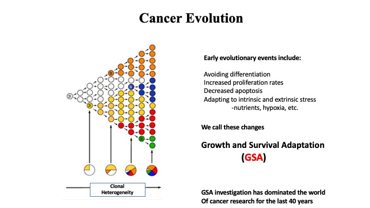
These alterations, which we will refer to as growth and survival adaptation (GSA), allow the tumor to escape the normal constraints of tissue homeostasis and to evolve into a relatively independent and distinctive tissue in its own right, with new rules and properties. Cancer research over the past 40 years has primarily occupied itself with understanding how genetic changes in the tumor allow these GSAs to come about.
Identification of cancer driver genes has traditionally proceeded along two main paths. The first was genetic and biochemical analyses looking either at viral oncogenes (1,2,3) or genes activated by viral insertions (4). The second involved identification of recurring mutations, first in familial cancer syndromes (5,6,7,8,9,10,11,12,13) and later sporadic cancers. (14,15,16) Modern analyses have streamlined these approaches by improved genetic approaches using transposons (17), RNA interference (RNAi), (18,19,20), CRISPR (21,22,23), and cDNA overexpression (24,25) together with breakthroughs in high-throughput sequencing to provide exhaustive lists of potential drivers. However, elucidation of how these genes function to drive cancer has lagged behind. Historically, systematic functional analysis of the genes responsible for GSA has been the main focus of cancer driver function, and many of the genetic screens have been limited to assessing functionality in traditional monoculture systems in vitro. These two-dimensional (2D) systems revealed genes required for tumor cell proliferation and survival but cannot account for the complexity of cell types and interactions that define the tumor microenvironment. Indeed, many known cancer driver mutations do not exhibit strong phenotypes in these 2D growth assays, raising the possibility that cancer-relevant phenotypes are being missed simply because of the assay system itself (26). For example, genetic screens in vivo using either RNAi or CRISPR have revealed phenotypes for putative squamous cell carcinoma drivers not appreciated in vitro (27,28).
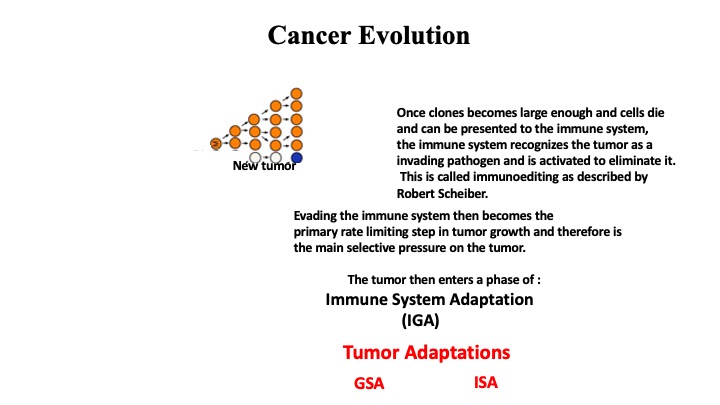
In addition to the GSAs that allow tumor emergence, there is another major barrier to overcome: the immune system. The immune system treats the tumor as an emerging pathogen and unleashes a response that aims to eliminate the tumor, thereby selecting for alterations that result in immune surveillance adaptation (ISA). Recently, in vitro CRISPR screens using co-cultures of isolated CD8 T cells and tumor cells have identified genes that regulate a tumor’s response to cytotoxic T cells (29), but these screens are predisposed toward uncovering genes involved in antigen presentation by major histocompatibility complex (MHC) class I and cannot account for other tumor and immune cell interactions found in vivo. The growing appreciation that immune evasion facilitates tumorigenesis has focused recent attention on this aspect of tumorigenesis and has led to revolutionary therapeutic approaches that overcome this evasion (30). Although immunoediting is a major mechanism of immune escape, several genes central to antigen presentation have been found to be recurrently mutated in cancer—for example, β2 microglobulin (B2M) and the human leukocyte antigen (HLA) alleles (31). In addition, a small number of cancer drivers such as KRAS, CDK4/6, and MYC have been implicated in aspects of immune evasion. However, the spectrum of genes involved in promoting immune recognition and their roles in cancer are not broadly known.
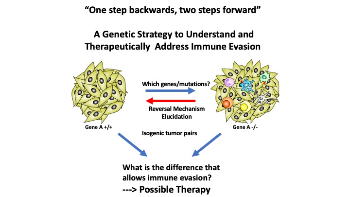
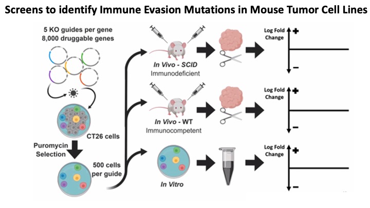
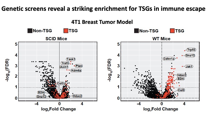
To approach this issue of immune evasion, we exploited syngeneic tumor models to perform CRISPR-based genetic screens in vitro and as tumor transplants in vivo in both immunocompetent [wild-type (WT)] and immunocompromised [Rag1null or severe combined immunodeficient (SCID)] mice that lack an adaptive immune system and uncovered a pronounced overlap between adaptive immune system–specific hits and the genes that are among the most frequently mutated in human tumors. In each tumor type tested, we found a marked enrichment for the loss of tumor suppressor genes (TSGs) in the presence of an adaptive immune system relative to immunocompromised mice. Nearly one-third of TSGs showed preferential enrichment, often in a cancer- and tissue-specific manner. These results suggest that clonal selection of recurrent mutations found in cancer is driven largely by the tumor’s requirement to avoid the adaptive immune system.
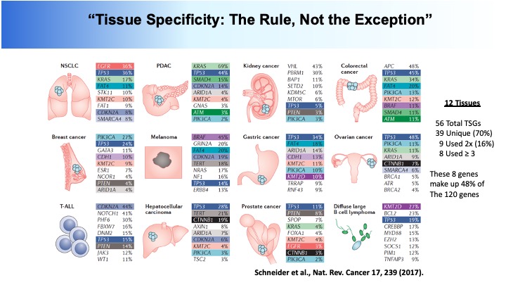
Profound tissue specificity in proliferation control underlies cancer drivers and aneuploidy patterns
Genomics has provided a detailed structural description of the cancer genome. Identifying oncogenic drivers that work primarily through dosage changes is a current challenge. Unrestrained proliferation is a critical hallmark of cancer. We constructed modular, barcoded libraries of human open reading frames (ORFs) and performed screens for proliferation regulators in multiple cell types. Approximately 10% of genes regulate proliferation, with most performing in an unexpectedly highly tissue-specific manner. Proliferation drivers in a given cell type showed specific enrichment in somatic copy number changes (SCNAs) from cognate tumors and helped predict aneuploidy patterns in those tumors, implying that tissue-type-specific genetic network architectures underlie SCNA and driver selection in different cancers. In vivo screening confirmed these results. We report a substantial contribution to the catalog of SCNA-associated cancer drivers, identifying 147 amplified and 107 deleted genes as potential drivers, and derive insights about the genetic network architecture of aneuploidy in tumors.
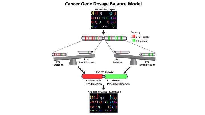
Cumulative haploinsufficiency and triplosensitivity drive aneuploidy patterns and shape the cancer genome
Aneuploidy has been recognized as a hallmark of cancer for more than 100 years, yet no general theory to explain the recurring patterns of aneuploidy in cancer has emerged. Here, we develop Tumor Suppressor and Oncogene (TUSON) Explorer, a computational method that analyzes the patterns of mutational signatures in tumors and predicts the likelihood that any individual gene functions as a tumor suppressor (TSG) or oncogene (OG).
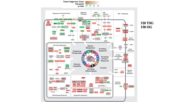
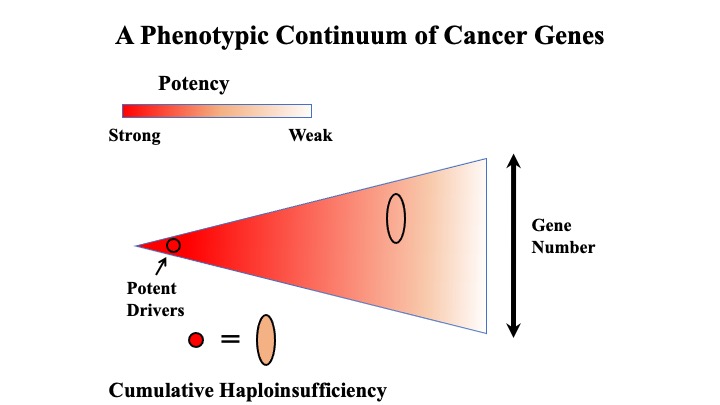
By analyzing >8,200 tumor-normal pairs, we provide statistical evidence suggesting that many more genes possess cancer driver properties than anticipated, forming a continuum of oncogenic potential.
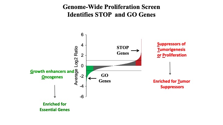
Integrating our driver predictions with information on somatic copy number alterations, we found that the distribution and potency of TSGs (STOP genes), OGs, and essential genes (GO genes) on chromosomes can predict the complex patterns of aneuploidy and copy number variation characteristic of cancer genomes. We propose that the cancer genome is shaped through a process of cumulative haploinsufficiency and triplosensitivity.
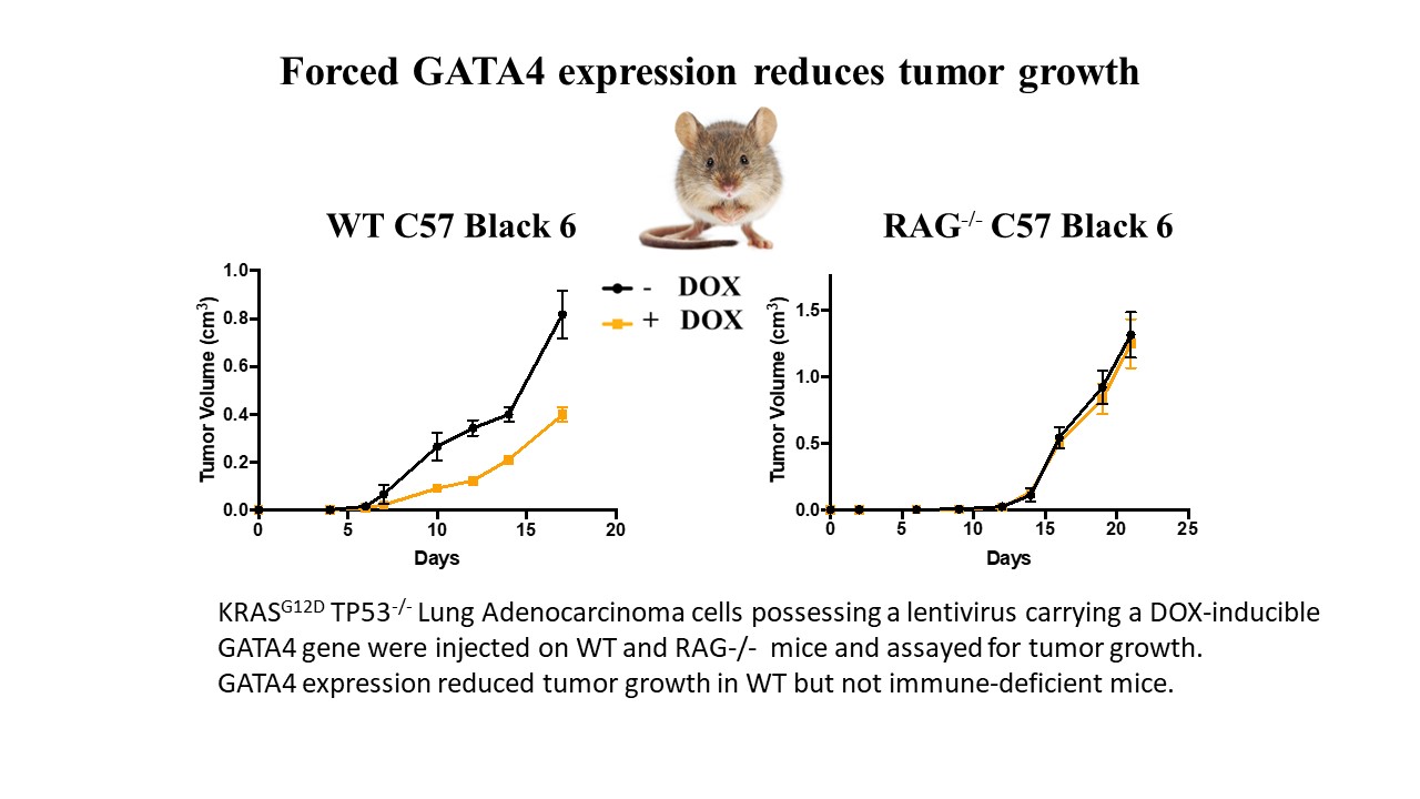
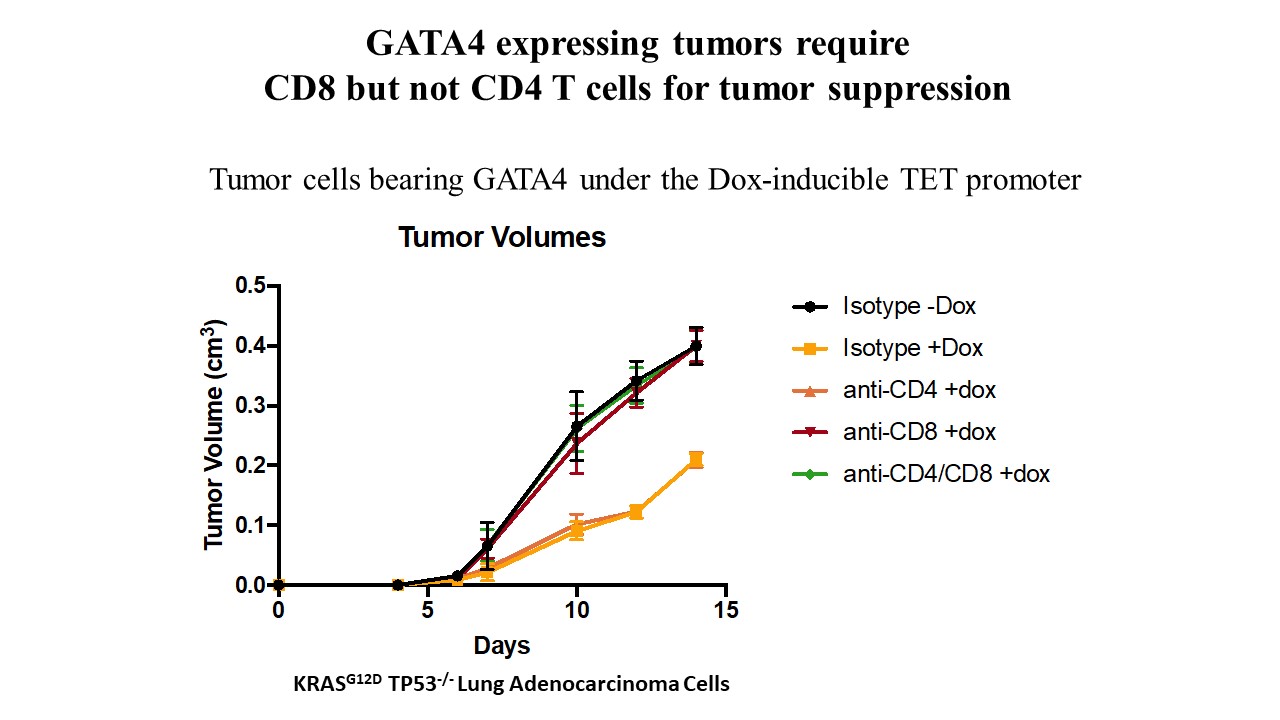
A GATA4-regulated secretory program suppresses tumors through recruitment of cytotoxic CD8 T cells
The GATA4 transcription factor acts as a master regulator of development of multiple tissues. GATA4 also acts in a distinct capacity to control a stress-inducible pro-inflammatory secretory program that is associated with senescence, a potent tumor suppression mechanism, but also operates in non-senescent contexts such as tumorigenesis. This secretory pathway is composed of chemokines, cytokines, growth factors, and proteases. Since GATA4 is deleted or epigenetically silenced in cancer, here we examine the role of GATA4 in tumorigenesis in mouse models through both loss-of-function and overexpression experiments. We find that GATA4 promotes non-cell autonomous tumor suppression in multiple model systems. Mechanistically, we show that GATA4-dependent tumor suppression requires cytotoxic CD8 T cells and partially requires the secreted chemokine Ccl2. Analysis of transcriptome data in human tumors reveals reduced lymphocyte infiltration in GATA4-deficient tumors, consistent with our murine data. Notably, activation of the GATA4-dependent secretory program combined with immune checkpoint inhibition with an anti-PD-1 antibody robustly abrogates tumor growth in vivo.
Genetic modifiers of the BRD4-NUT dependency of NUT midline carcinoma uncovers a synergism between BETi and CDK4/6i inhibitors
Bromodomain and extraterminal (BET) domain inhibitors (BETis) show efficacy on NUT midline carcinoma (NMC). However, not all NMC patients respond, and responders eventually develop resistance and relapse. Using CRISPR and ORF expression screens, we systematically examined the ability of cancer drivers to mediate resistance of NMC to BETis and uncovered six general classes/pathways mediating resistance. Among these, we showed that RRAS2 attenuated the effect of JQ1 in part by sustaining ERK pathway function during BRD4 inhibition. Furthermore, overexpression of Kruppel-like factor 4 (KLF4), mediated BETi resistance in NMC cells through restoration of the E2F and MYC gene expression program. Finally, we found that expression of cyclin D1 or an oncogenic cyclin D3 mutant or RB1 loss protected NMC cells from BETi-induced cell cycle arrest. Consistent with these findings, cyclin-dependent kinase 4/6 (CDK4/6) inhibitors showed synergistic effects with BETis on NMC in vitro as well as in vivo, thereby establishing a potential two-drug therapy for NMC. This therapy has now entered a clinical trial.
A genetic interaction analysis identifies cancer drivers that modify EGFR dependency
A large number of cancer drivers have been identified through tumor sequencing efforts, but how they interact and the degree to which they can substitute for each other have not been systematically explored. To comprehensively investigate how cancer drivers genetically interact, we searched for modifiers of epidermal growth factor receptor (EGFR) dependency by performing CRISPR, shRNA, and expression screens in a non-small cell lung cancer (NSCLC) model. We elucidated a broad spectrum of tumor suppressor genes (TSGs) and oncogenes (OGs) that can genetically modify proliferation and survival of cancer cells when EGFR signaling is altered. These include genes already known to mediate EGFR inhibitor resistance as well as many TSGs not previously connected to EGFR and whose biological functions in tumorigenesis are not well understood. We show that mutation of PBRM1, a subunit of the SWI/SNF complex, attenuates the effects of EGFR inhibition in part by sustaining AKT signaling. We also show that mutation of Capicua (CIC), a transcriptional repressor, suppresses the effects of EGFR inhibition by partially restoring the EGFR-promoted gene expression program, including the sustained expression of Ets transcription factors such as ETV1. Together, our data provide strong support for the hypothesis that many cancer drivers can substitute for each other in certain contexts and broaden our understanding of EGFR regulation.
SPECIFICANCER: A Cancer Grand Challenges Research Consortium
In a collaboration that involves researchers from multiple disciplines – including geneticists, cell biologists and bioinformaticians – this Grand Challenge project aims to generate a comprehensive map of cancer drivers and their specificity to different tissues. This has the potential to improve our basic understanding of cancer, and provide information that will impact therapeutic choices for patients.
Our international team is made up of researchers from the US, the UK, and the Netherlands.
We are going to look at our cells’ DNA to identify which genes control whether cells divide or not. By looking at the DNA in different locations in the body, this screen will allow the team to look at whether certain genes are only active in specific tissues. This will provide vital information on known cancer drivers, as well as allowing the team to identify new potential drivers of cancer. Alongside this, the researchers will be assessing how well different cancer drugs work in different types of cancer, and if this can be linked to the activity of cancer drivers.
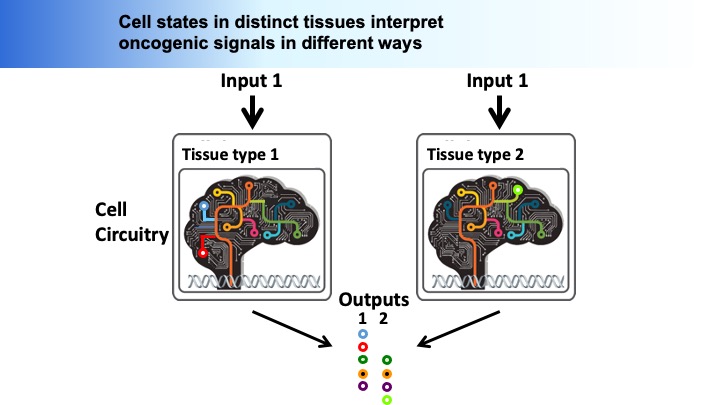
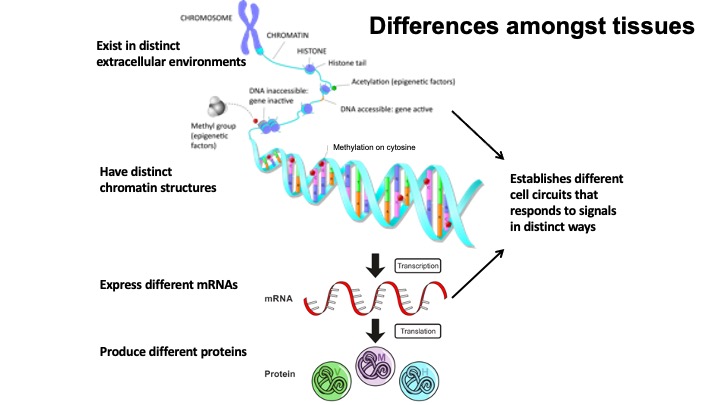
Central to SPECIFICANCER’s hypothesis is that cancer develops differently in different organs because of the way they are programmed – for example, a brain cell has a very different function to a skin cell. In both cases, these cells possess the same DNA, but the DNA is organised into a tissue-specific network, dictating a very specific behaviour and function. Understanding how this DNA is programmed is key to understanding the tissue-specifics of cancer.
The team is driving a range of approaches to understand this – one of which is to home in on basic biology. By scrutinising healthy cells from the 8 tissue types that give rise to the most common cancers – breast, bowel, lung, skin, kidney, liver, brain and pancreas – the team hopes to identify whether certain genes are only active in different parts of the body. They’ll also introduce mutations into hundreds of genes to see which ones drive cancer in the different tissue types.
If successful, this map of ‘tissue specificity’ will give us a complete overview of which cancer drivers play a role in the different tissues throughout the body. This understanding could transform the way doctors treat cancer, as they will be able to select which drugs are more likely to work based on exactly how and where the cancer originated.
Older Work
Genetic identification of tumor suppressors: isolation of REST, INNP4
Tumorigenesis is a multistep process characterized by a myriad of genetic and epigenetic alterations. Identifying the causal perturbations that confer malignant transformation is a central goal in cancer biology. To identify genes involed in tumorigenesis we performed an RNAi-based genetic screen for genes that suppress transformation of human mammary epithelial cells. We identified several genes previously implicated in proliferative control and epithelial cell function including two established tumor suppressors, TGFBR2 and PTEN (181). In addition, we uncovered a previously unrecognized tumor suppressor role for REST/NRSF, a transcriptional repressor of neuronal gene expression. Array-CGH analysis identified REST as a frequent target of deletion in colorectal cancer. Furthermore, we detect a frame-shift mutation of the REST gene in colorectal cancer cells that encodes a dominantly acting truncation capable of transforming epithelial cells. Cells lacking REST exhibit increased PI(3)K signaling and are dependent upon this pathway for their transformed phenotype. These results implicate REST as a human tumor suppressor and provide a novel approach to identifying candidate genes that suppress the development of human cancer.
We also identified a novel component of the PI3K pathway, PI(3,4)P2 phosphatase INPP4B. Loss of INPP4 allows cells to grow in an anchorage-independent manner. This finding was further examined by Lew Cantley’s group, who found that INPP4 controls inositol phosphate pathways in vivo and is deleted in tumors. These screens were performed with our generation 1 shRNA libraries. These screens are now being repeated with our second generation microRNA shRNA libraries.
The tumor suppressor REST is ubiquitinated by the oncogene complex SCFBTRCP
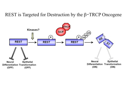
The RE1-silencing transcription factor (REST, also known as NRSF) is a master repressor of neuronal gene expression and neuronal programmes in non-neuronal lineages. We identified REST as a human tumour suppressor in epithelial tissues, suggesting that its regulation may have important physiological and pathological consequences. However, the pathways controlling REST had yet to be elucidated. We found that REST is regulated by ubiquitin-mediated proteolysis and used an RNA interference (RNAi) screen to identify a Skp1-Cul1-F-box protein complex containing the F-box protein BTRCP (SCFBTRCP) as an E3 ubiquitin ligase responsible for REST degradation. SCFBTRCP binds and ubiquitinates REST and controls its stability through a conserved phospho-degron (208). During neural differentiation, REST is degraded in a beta-TRCP-dependent manner. BTRCP is required for proper neural differentiation only in the presence of REST, indicating that BTRCP facilitates this process through degradation of REST. Conversely, failure to degrade REST attenuates differentiation. Furthermore, we find that BTRCP overexpression, which is common in human epithelial cancers, causes oncogenic transformation of human mammary epithelial cells and that this pathogenic function requires REST degradation. Thus, REST is a key target in BTRCP-driven transformation and the BTRCP-REST axis is a new regulatory pathway controlling neurogenesis. We also showed that a REST-associated protein, CDYL, is also required to suppress cellular transformation in HMECs (220).
Genetic screening for cancer lethal genes
A key for the discovery of anticancer drug targets is the identification of genes upon which cancer cells rely for survival. Although we know the identity of a number of oncogenes upon which tumors depend, there is likely many more genes cancer cells need for their proliferation and survival. Unfortunately because cancer cells have extensively rewired their cellular circuitry, it is not possible to predict cellular dependencies. An unbiased method for identifying such dependencies is through genetic screening. We have developed highly parallel multiplex methodology for screening large pools of shRNAs using half-hairpin barcodes for microarray deconvolution that allows genetic screens to be performed to identify growth promoters and growth inhibitors.
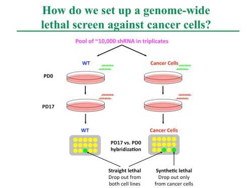
We carried out dropout screens for shRNAs that affect cell proliferation and viability in cancer cells and normal cells (205, 206). We identified many shRNAs to be antiproliferative that target core cellular processes, such as the cell cycle and protein translation, in all cells examined. Moreover, we identified genes that are selectively required for proliferation and survival in different cell lines which suggests that this approach can be employed to identify new cancer drug targets.
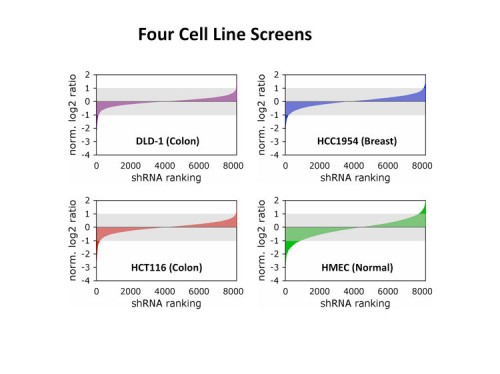
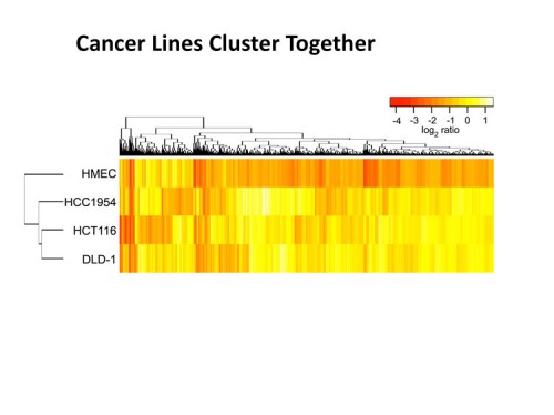
Our platform enables rapid and cost-effective genome-wide screens to identify cancer proliferation and survival genes for target discovery. Such efforts are complementary to the Cancer Genome Atlas and provide an alternative functional view of cancer cells. Among the discoveries made using this preliminary analysis was the fact that it is much easier to identify genes that control growth in normal cells than in cancer cells. This is the unfortunate dilemma of drug discovery, normal cells are killed much more easily than cancer cells. However, we did discover that there are cancer-specific lethals and that cancer cells have a lethality signature that is closer to other cancer cells than normal cells.
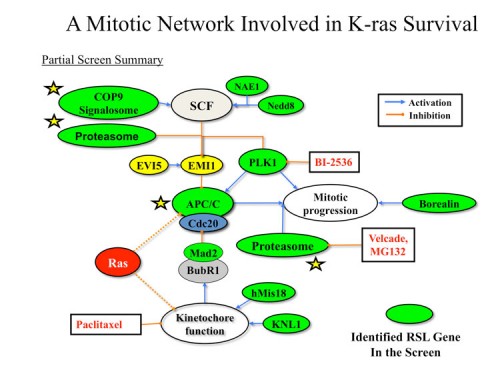
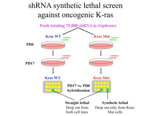
Synthetic lethal screening with the human oncogenic mutations in KRAS
It is relatively simple to identify cancer lethal shRNAs but it is important to understand why they are killing the cancer cells if we ever wish to employ a strategy to identify which cancers to apply a particular therapy towards. For this reason instead of searching for shRNAs that kill cancer cell lines alone, we have begun to identify shRNAs that kill cancer cells that carry a particular oncogenic lesion. We first focused on identifying synthetic lethal interaction with the Ras oncogene using our genome-wide barcoded shRNA library (228). This approach uncovered a diverse set of genes whose knockdown selectively impairs the viability of Ras mutant cells. We identified a concentration of genes involved in mitosis. Among these are the mitotic kinase PLK1 and the anaphase promoting complex/cyclosome. Gene expression analysis indicates that reduced expression of genes in this pathway also correlates with increased survival of patients bearing tumors with a mutant Ras transcriptional signature. Thus, targeting this pathway might be useful for the treatment of Ras-driven cancers. Importantly, many of the Ras synthetic lethal genes we identified in this screen are not mutated or themselves oncogenes. We therefore propose a concept of “non-oncogene addiction” to describe this addiction of cancer cells to the function of normal cellular genes to alleviate aspects of oncogenic stress (see our review in 2009 Cell “Principles of Cancer Therapy” (228)). Like oncogene-addiction, drugs targeting non-oncogene addiction should provide a valuable approach for the treatment of cancer.
Identification of the PTPN12 tyrosine phosphatase as a tumor suppressor
Among breast cancers, triple-negative breast cancer (TNBC) is the most poorly understood and is refractory to current targeted therapies. Using a genetic screen for shRNAs that suppress cellular transformation (growth in soft agar), we identified the PTPN12 tyrosine phosphatase as a tumor suppressor in TNBC. PTPN12 potently suppresses mammary epithelial cell proliferation and transformation. PTPN12 is frequently compromised in human TNBCs, and we identify an upstream tumor-suppressor network that posttranscriptionally controls PTPN12. PTPN12 suppresses transformation by interacting with and inhibiting multiple oncogenic tyrosine kinases, including HER2 and EGFR. The tumorigenic and metastatic potential of PTPN12-deficient TNBC cells is severely impaired upon restoration of PTPN12 function or combined inhibition of PTPN12-regulated tyrosine kinases, suggesting that TNBCs are dependent on the proto-oncogenic tyrosine kinases constrained by PTPN12. Collectively, these data identify PTPN12 as a commonly inactivated tumor suppressor and provide a rationale for combinatorially targeting proto-oncogenic tyrosine kinases in TNBC and other cancers based on their profile of tyrosine-phosphatase activity.
Review: Current State of Cancer Research
Principles of cancer therapy: oncogene and non-oncogene addiction
The past two decades have witnessed tremendous advances in our understanding of the pathogenesis of cancer. It is now clear that cancer arises through a multi-step, mutagenic process whereby cancer cells acquire a common set of properties including unlimited proliferation potential, self-sufficiency in growth signals, and resistance to anti-proliferative and apoptotic cues. Furthermore, tumors evolve to garner support from surrounding stromal cells, attract new blood vessels to bring nutrients and oxygen, evade immune detection, and ultimately metastasize to distal organs. Many of these phenotypic traits can be brought about by genetic alterations that involve the gain-of-function mutation, amplification, and/or over-expression of key oncogenes together with the loss-of-function mutation, deletion, and/or epigenetic silencing of key tumor suppressors
Cancer cells achieve these phenotypes in large part by reactivating and modifying many existing cellular programs normally used during development. These programs control coordinated processes such as cell proliferation, migration, polarity, apoptosis and differentiation during embryogenesis and tissue homeostasis. Consistent with Darwinian principles, cancer evolves through random mutations and epigenetic changes that alter these pathways followed by the clonal selection of cells that can survive and proliferate under circumstances that would normally be deleterious.
Although a number of oncogenes and tumor suppressors are frequently mutated in cancer cells, such as PI3K, Ras, p53, PTEN, Rb, and p16INK4a, there also appears to be a large number of low frequency changes that can contribute to oncogenesis. Indeed, data from tumor sequencing projects reveal an astounding diversity of mutations in tumors. In one study, Stratton and colleagues estimate that individual mutations in as many as 20% of all kinases can play an active role in tumorigenesis (Greenman et al., 2007), although it remains to be seen whether mutations in 20% of other gene classes will also drive tumorigenesis. Large-scale sequencing of multiple cancers have so far failed to identify new, high-frequency mutation targets in addition to those previously identified (Cancer Genome Atlas Research Network, 2008; Ding et al., 2008; Jones et al., 2008; Parsons et al., 2008; Sjoblom et al., 2006; Wood et al., 2007). Rather, these studies found that every tumor harbors a complex combination of low-frequency mutations thought to drive the cancer phenotype. Furthermore, the repertoires of somatic mutations in different cancer types such as breast and colon cancers appear to be different. Although there is much debate with regard to the statistical requirements needed to distinguish likely driver from non-contributing passenger mutations among the large collection of mutations in tumors, it is clear that there is tremendous complexity and heterogeneity in the patterns of mutations in tumors of different origins.
The complexity of alterations in cancer presents a daunting problem with respect to treatment: how can we effectively treat cancers arising from such varied perturbations? Cancer cells have extensively rewired pathways for growth and survival that underlie the malignant phenotype. Thus, a key to successful therapy is the identification of critical, functional nodes in the oncogenic network whose inhibition will result in system failure, that is, the cessation of the tumorigenic state by apoptosis, necrosis, senescence, or differentiation. Furthermore, therapeutic agents attacking these nodes must display a sufficiently large therapeutic window with which to kill tumor cells while sparing normal cells. To borrow a term from yeast and fly genetic analyses, the therapeutic agents must constitute “synthetic lethality” with the cancer genotype/phenotype. In some cases, particular agents can display genotype-dependent lethality similar to synthetic lethality without directly inhibiting a particular protein. The efficacy of the two mainstay treatment options for cancer today—chemotherapy and radiation—are examples of agents that exploit the enhanced sensitivity of cancer cells to DNA damage. Despite all of our knowledge, however, we still do not have a clear molecular understanding of why these agents work to selectively kill tumor cells, and, conversely, why they eventually fail. The advent of “targeted” therapies, which aim to attack the underlying oncogenic context of tumors, provides more sophisticated examples of synthetic lethality. When properly deployed, these therapies tend to be more effective relative to chemotherapy and radiation.
The hallmarks of cancer
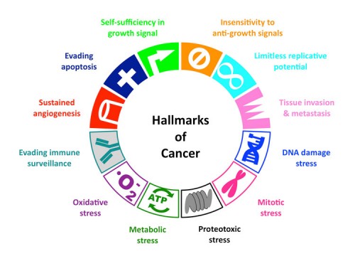
Although there is no simple way to predict a priori which proteins will act as nodal points to generate cancer drug targets, solutions are likely to emerge from multiple sources, including recent initiatives to understand cancer at the systems level. From a genetic point of view, it is important to appreciate that the plethora of mutations observed in the cancer genome must ultimately result in a common set of hallmarks in order to bring about the malignant phenotype. The goal of cancer therapy is, therefore, to either reverse these properties or target them as tumor-specific liabilities, preferably through the combinatorial application of a relatively small number of drugs. Thus we need a thorough understanding of the nature of these hallmarks.
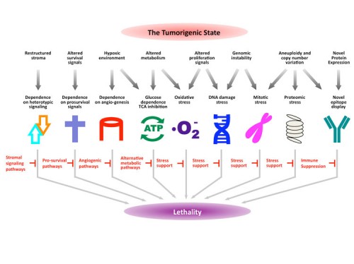
In addition to the six hallmarks outlined in the seminal review by Hanahan and Weinberg (Hanahan and Weinberg, 2000) that collectively promote survival and proliferation in foreign environments as well as the hallmark of “evading immune surveillance” proposed by Kroemer and colleagues (Kroemer and Pouyssegur, 2008) we propose a number of additional, equally prevalent hallmarks of cancer cells based on recent analyses of cellular phenotypes. Although these cancer phenotypes are not responsible for initiating tumorigenesis, they are common characteristics of many tumor types. Among these additional hallmarks are DNA damage/replication stress, proteotoxic stress, mitotic stress, metabolic stress, and oxidative stress. We collectively refer to this subset as the stress phenotypes of cancers. There are often intricate functional interplays among these shared hallmarks of tumor cells, which are illustrated and discussed below. Although some of these stress phenotypes are not unique to cancer cells and can be observed in other conditions such as chronic inflammation, we propose that they represent a common set of oncogenesis-associated cellular stresses that cancer cells must tolerate through stress support pathways. How these phenotypes arise is not well understood, but targeting these hallmarks and their associated vulnerabilities in a wide variety of cancers has shown promise for therapeutic intervention.
DNA damage and DNA replication stress
Based on karyotypic and mutational analyses, it is clear that tumors, especially solid tumors, pass through stages of extreme genomic instability that result in the accumulation of point mutations, deletions, complex chromosomal rearrangements and extensive aneuploidy (Hartwell and Kastan, 1994). This level of instability is due in part to a constitutive level of endogenous DNA damage, which results in activation of the DNA damage stress response (DDR) pathway (Bartkova et al., 2005; Gorgoulis et al., 2005). Elevated levels of DNA damage observed in early stage tumors are thought to be due to several factors. First, the shortening of telomeres due to replication in the absence of sufficient telomerase activity leads to the appearance of double strand breaks (DSBs) at telomeric ends. The subsequent fusions of these de-protected ends initiate breakage-fusion-bridge cycles that result in translocations and gene amplification events (Maser and DePinho, 2002). DSBs resulting from replication stress can also lead to breakage-fusion-bridge cycles (Windle et al., 1991). Additionally, oncogene activation in pre-cancerous lesions has been shown to increase DSBs and genomic instability (Halazonetis et al., 2008), possibly through DNA hyper-replication (Bartkova et al., 2006; Di Micco et al., 2006). Finally, mutation of genes involved in either DNA repair programs (such as excision, cross-link or mismatch repair) or the DDR pathways (such as ATM and p53 signaling), can lead to increased DNA damage, inappropriate cell cycle progression and genomic instability (Harper and Elledge, 2007). In normal cells, DNA damage signals to halt proliferation, induce cellular senescence, or elicit apoptosis. Cancer cells have evolved to overcome the anti-proliferative effects of DNA damage, continuing to replicate in the presence of damage.
Proteotoxic stress
Tumors exhibit proteotoxic stress evidenced by their frequent constitutive activation of the heat shock response. We think this is due, in part, to the striking degree of aneuploidy (altered chromosome number) often found in solid tumors (see figure below) (Ganem et al., 2007; Torres et al., 2008; Williams et al., 2008). Aneuploidy and gene copy-number changes can alter the relative balance of growth and survival signals, thereby promoting tumorigenesis. However, they also result in corresponding increases and decreases in transcript levels (Pollack et al., 2002; Torres et al., 2007; Tsafrir et al., 2006) that produce imbalances in the stoichiometry of protein complex subunits (Papp et al., 2003). These imbalances increase the amount of toxic, unfolded protein aggregates in the cell and place additional burdens on the protein folding and degradation machineries (Denoyelle et al., 2006). This proteotoxic stress is counteracted, in part, by the heat shock response pathway, which promotes the proper folding and/or proteolytic degradation of proteins (Whitesell and Lindquist, 2005).
Mitotic stress
A subset of tumors display increased rates of chromosome mis-segregation, which is referred to as the CIN (chromosome instability) phenotype (Komarova et al., 2002). This instability results in a shifting chromosome distribution, thus allowing tumor cells to rapidly evolve. In principle, CIN phenotypes can result from defects in a variety of pathways involved in mitosis, including defects in mitotic proteins that execute chromosome segregation and defects in the spindle assembly checkpoint, which coordinates anaphase entry with proper alignment of chromosomes on the mitotic spindle (Cahill et al., 1998). In addition, the CIN phenotype could result from the presence of extra centrosomes in tumor cells or from stresses placed on the mitotic apparatus due to the need to segregate supernumerary chromosomes (Ganem et al., 2007). Furthermore, CIN and mitotic stress might arise indirectly as a result of DSBs and genomic instability following oncogene activation, even in lesions where the mitotic machinery is intact (Halazonetis et al., 2008). Mutations in certain oncogenes, such as Ras, and tumor suppressors, such as p53, have been suggested to contribute to the CIN phenotype (Denko et al., 1994). However, the precise cause of mitotic stress is not known for the vast majority of tumors.
Metabolic stress
Normal cells derive the bulk of their ATP through mitochondrial oxidative phosphorylation. In what has been referred to as the Warburg effect, most cancer cells are found to predominantly produce energy by the less efficient method of glycolysis and secrete a large amount of lactic acid, even under high oxygen conditions (Warburg, 1956). Tumor cells exhibit dramatically increased glucose uptake and highly elevated rates of glycolysis (DeBerardinis et al., 2007). This provides the basis for tumor imaging by positron emission tomography (PET) using the glucose analog 18F-2-deoxyglucose. This transition to glycolysis for energy production provides several advantages to the tumor including adaptation to a low oxygen environment and the acidification of the surrounding microenvironment, which promotes tumor invasion and suppresses immune surveillance.
Oxidative stress
The defining characteristic of oxidative stress is the presence of reactive oxygen species (ROS) and cancer cells typically generate more ROS than normal cells (Szatrowski and Nathan, 1991). Both oncogenic signaling (Lee et al., 1999) and the down-regulation of mitochondrial function (Gogvadze et al., 2008) in tumors can contribute to ROS generation. ROS are highly reactive and likely to contribute to the increased levels of endogenous DNA damage observed in cancer cells. In addition, ROS are important signaling mediators, and their presence may contribute to transformation. For example, ROS promote the activation of the transcription factor HIF-1 by hypoxia (Dewhirst et al., 2008), and HIF-1 can promote the glycolytic switch andgenesis observed in tumors.
Attacking the hallmarks of cancer
Any therapy with the stated goal to treat and possibly cure cancer must show differential toxicity towards tumor cells relative to normal cells. Implicit in this statement is that some unique properties of cancer cells not shared by normal cells, such as those depicted in Figure 1, must be exploited to the specific detriment of cancer cells, i.e. the concept of synthetic lethality. In principle, cancer can be treated by inducing cancer cells to undergo apoptosis, necrosis, senescence, or differentiation. These changes can be brought about by disrupting cancer cell-autonomous processes, by interfering with autocrine/paracrine signaling within tumors, or by blocking heterotypic signaling between tumor cells and the surrounding stromal tissue or blood vessels. Enhancing immune surveillance against cancer cells expressing novel antigens is also an attractive approach that has shown efficacy in specifically killing cancer cells (Muller and Scherle, 2006).
Experiments aimed at either suppressing oncogene activity or restoring tumor suppressor function have revealed that such intervention is highly deleterious to the cancer cell. The heightened state of dependency of cancer cells on oncogenes and the loss of tumor suppressors led to the terms “oncogene addiction” (OA) and “tumor suppressor gene hypersensitivity” (Weinstein, 2002; Weinstein and Joe, 2008). These alterations are necessary for both the establishment and maintenance of the oncogenic state and therefore are logical drug targets. Indeed, much effort has been extended to pharmacologically inhibit oncoproteins. What is thought to underlie the phenomenon of oncogene addiction is the observation that oncogenes elicit strong, opposing pro-survival and pro-apoptotic signals in cancer cells that favor growth and survival, and the acute inhibition of oncogene function tilts this balance towards cell death (Downward, 2003; Sharma and Settleman, 2007).
To bring about their phenotypic manifestations, oncogenes rely on extensive adaptations in cellular processes that are themselves not oncogenic. In addition, cancer cells may also display an increased dependence on the normal cellular functions of certain genes that act in oncogenic pathways but are not themselves classical oncogenes. For example, mutations in many genes in a given oncogenic pathway are unable to directly promote tumor formation because, despite being required for their pathway, they cannot increase the overall activity of the pathway because they are not rate-limiting. However, a reduction in the activity of many such genes can become rate-limiting to the pathway in question and, thus, they represent potential drug targets. By this rationale, cancer cells are addicted to both oncogenes and non-oncogenes. To describe this addiction of cancer cells to the functions of non-oncogenes, we have termed this phenomenon “non-oncogene addiction”, NOA (Solimini et al., 2007). Although NOA genes, like oncogenes, are required for maintenance of the tumorigenic state, NOA genes do not undergo oncogenic mutations or functionally significant genomic alterations in tumors. The concept of non-oncogene addiction underscores the important contribution of these supporting networks to oncogenesis and highlights the potential of non-oncogenes as points of intervention for cancer therapeutics.
Whereas some gene classes and pathways fall neatly into the OA or NOA designations, others are more difficult to categorize because they exhibit characteristics of both phenomena. For example, interferon regulatory factor 4 (IRF4) (Iida et al., 1997) is oncogenic and over-expressed due to translocations in some multiple myelomas. However, it is also required for the survival of myelomas lacking IRF4 translocations or overexpression (Shaffer et al., 2008). Should it be considered as an example of OA in the latter cases? Also, should a protein that is directly activated by an oncogene and required for tumorigenesis – but is otherwise not mutated in cancer – be considered an example of NOA when it is so clearly linked to an oncogene? Both examples are clear if one adheres to a strict definition of NOA stating that NOA genes do not undergo oncogenic mutations in tumors. However, these examples often run counter to our overall intuitive sense of the different categories. Regardless, although the OA and NOA designations are not perfect, they provide a useful intellectual framework for thinking about cancer cell vulnerabilities and the principles of cancer therapies. Below, we will discuss examples of oncogene and non-oncogene addiction and describe how modern tools are being applied to identify these classes of genes for possible therapeutic exploitation.
Oncogene addiction and tumor suppressor gene hypersensitivity
Despite the multitude of genetic and epigenetic alterations found across cancers, a given tumor is likely to be driven by only a select few changes—those that result in the gain of an oncogene or the loss of a tumor suppressor. The phrase “oncogene addiction” was coined to describe the observation that tumor maintenance often depends upon the continued activity of certain oncogenes (Weinstein, 2002). This phenomenon has been demonstrated in vivo for several oncogenes. For example, mouse models using an inducible MYC oncogene have shown that MYC-driven skin papillomas, lymphomas, and osteosarcomas can all be reversed upon MYC withdrawal (Felsher and Bishop, 1999; Jain et al., 2002; Pelengaris et al., 1999). Similarly, addictions to the HRAS or BCR-ABL oncogenes have been demonstrated in mouse models of melanoma and leukemia, respectively (Chin et al., 1999; Huettner et al., 2000). In human colorectal cancer cells bearing a KRAS mutation, somatic knockout of the KRAS oncogene results in reversion of the transformed phenotype and abrogates the ability of these cells to form tumors in nude mice (Shirasawa et al., 1993).
The subset of oncogenes whose inhibition can lead to tumor cell death, differentiation, arrest, or senescence is of great clinical interest as targets for cancer therapeutics This strategy has proven successful for the protein kinase oncogenes BCR-ABL (imatinib/Gleevec), EGFR (gefitinib/Iressa, erlotinib/Tarceva), and HER2 (trastuzumab/Herceptin) (Druker, 2002; Roberts and Der, 2007; Sharma et al., 2007) and efforts towards inhibition of BRAF, MDM2 and the lipid kinase PI3K are underway. Targeting non-kinase oncogenes such as RAS and MYC, however, has proven more difficult.
In contrast to oncogenes, tumor suppressor genes act to provide the cellular restraints necessary to prevent aberrant growth and survival or genomic instability. Loss of tumor suppressor genes through deletion, inactivating mutation, or epigenetic silencing results in the removal of these restraints leading to tumorigenesis. Reintroduction of a tumor suppressor gene into a tumor lacking that gene can result in tumor regression. This concept has been recently demonstrated by reactivation of p53 in mouse tumor models (Martins et al., 2006; Ventura et al., 2007; Xue et al., 2007). Pharmacological exploitation of tumor suppressor mutations, however, have lagged behind efforts aimed at oncogenes because it is often difficult to use a small molecule to either restore or mimic the function of a protein that is either mutated or absent. In cases where a tumor suppressor negatively regulates the activity of a proto-oncogene, drugs targeting the corresponding proto-oncogene should prove efficacious in treating tumors lacking that tumor suppressor. For example, tumors that have lost the tumor suppressor and lipid phosphatase PTEN, which normally acts to constrain PI3K signaling, are likely to be sensitive to PI3K inhibitors. Similarly, loss of Rb, p16, p21, or p27 all result in up-regulation of cyclin-dependent kinase (CDK) activity, which drives cell cycle entry. In principle, tumors resulting from these lesions might be more sensitive to CDK inhibitors. Whether such predictions prove true will only become apparent from clinical trials employing PI3K and CDK inhibitors. In many other cases, however, such as those involving loss of the tumor suppressors p53 or ARF, there is no obvious positive signaling pathway to target, and alternative therapeutic strategies must therefore be considered.
Non-oncogene addiction: a theoretical framework for targets for cancer therapy
We proposed the concept of non-oncogene addiction (NOA) based on the understanding that the tumorigenic state depends on the activities of a wide variety of genes and pathways, many of which are not inherently oncogenic themselves (Solimini et al., 2007). Importantly, these genes and pathways are essential to support the oncogenic phenotype of cancer cells, but are not required to the same degree for the viability of normal cells. From a purely genetic point of view, these dependencies should provide an ample number of drug targets that when inhibited will constitute synthetic lethality with the underlying tumor genotype. Gene interaction studies in yeast have provided precedence for this notion. For example, most mutations exhibit enhanced growth defects when paired with certain other mutations, and one study in yeast identified an average of six genetic interactions per gene (Collins et al., 2007). As a tumor contains many genetic alterations, each of these changes provides an opportunity to pair with the loss of function of a second gene to result in a severe and possibly lethal growth and survival phenotype. Furthermore, if this second gene is targeted with a drug that inhibits its protein, then a potential cancer therapy can result.
NOA genes and pathways provide important targets for anti-tumor therapies. In the sections below, we will discuss general classes of NOA genes with specific examples when available. NOA genes fall into two general categories, tumor intrinsic and tumor extrinsic. Whereas tumor intrinsic NOA genes support the oncogenic state of the tumor cell in a cell-autonomous manner, tumor extrinsic NOA genes function in stromal and vascular cells that provide heterotypic support for the tumor. An advantage of targeting these accessory cells is that, unlike tumor cells, they tend to be genetically more stable and therefore are less likely to evolve drug resistance. However, in certain circumstances tumors may be able to evolve reduced dependency on these accessory cells.
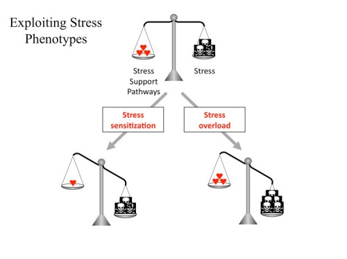
Intrinsic non-oncogene addiction
The trade-off for the tumorigenic state is that tumor cells experience numerous cellular stresses not experienced by normal cells and therefore tumor cells are more dependent on stress support pathways for their survival. In principle, there are two approaches to exploit this dependency to selectively kill tumor cells. The first approach, stress sensitization, aims to diminish the activity of the stress support pathways such that the tumor cell can no longer cope with the stress of its oncogenic state and either ceases to proliferate or initiates apoptosis or necrosis. The second approach, stress overload, aims to exacerbate existing oncogenic stress in order to overwhelm the stress support pathways in the tumor cell, leading to growth arrest or cell death. Both approaches, therefore, disrupt the balance of pro- and anti-survival signaling to the detriment of tumor cells. Examples of specific types of NOA are illustrated and discussed below.
DNA damage and replication stress
In nearly all tumor cell types, the rate of spontaneous DNA damage and the degree of replication stress are enhanced. The presence of oncogenes can also elicit substantial DNA damage (Halazonetis et al., 2008). DNA damage stress can be exploited therapeutically through both stress sensitization and stress overload. An elaborate DNA damage response (DDR) pathway exists in the cell to ameliorate the effects of DNA damage by promoting DNA repair. Mutations in genes of this pathway result in increased sensitivity to DNA damage (Harper and Elledge, 2007). In principle, cells experiencing spontaneous DNA damage should show enhanced sensitivity to agents that interfere with this stress response pathway In support of this notion, recent studies have shown that inhibitors of the DDR kinases ATM and Chk1 exhibit selective toxicity towards cancer cells (Chen et al., 2006; Kennedy et al., 2007). Although it seems counter-intuitive that a pathway that normally serves to restrain proliferation and promote apoptosis would protect a cancer cell, the DNA repair and genomic stability afforded by this pathway could save a cancer cell from death caused by persistent DNA damage and replication stress.
Stress overload of the DDR pathway should also show efficacy in cancer treatment. Although it is not clear why DNA damaging agents, such as IR and chemotherapy, are effective cancer therapies, it is possible that these are examples of stress overload, where cancer cells with already elevated levels of DNA damage and replication stress cannot repair the additional damage inflicted by these agents. An alternative explanation is that during tumorigenesis, the persistence of DNA damage selects for cells with mutations that abrogate part of the DDR pathway and therefore cannot properly sense and respond to DNA damage. These cells with a partially defective DDR might therefore be more vulnerable to the extensive DNA damage resulting from radiation or chemotherapy that is lethal without a normal DDR pathway. In this context, DNA damage exploits a stress phenotype of tumors that is analogous to non-oncogene addition.
Given the sensitivity of many cancers to DNA damaging agents, there should exist genes whose inhibition will generate endogenous DNA damage to exacerbate this sensitivity. Exemplifying this phenomenon are cancers bearing BRCA2 mutations. These tumors are defective in homologous recombination-mediated DNA repair and are particularly sensitive to DNA cross-linkers such as cisplatin (Sakai et al., 2008). Furthermore, these tumors rely heavily on other forms of DNA repair such as base-excision repair, resulting in sensitivity to inhibitors of poly-ADP-ribose polymerase (PARP1), an enzyme that facilitates repair of single stranded breaks (Bryant et al., 2005; Farmer et al., 2005). In normal cells, the endogenous DNA damage generated by PARP inhibition is well-tolerated because of functional compensation from homologous recombination-mediated repair. Indeed, mice deficient in PARP1 are fertile and do not exhibit increased tumor susceptibility (Conde et al., 2001). The addiction of BRCA2-mutant cells to PARP1 function is therefore an example of NOA that is exploited therapeutically. There are likely to be additional non-oncogene targets like PARP1 that can be exploited to treat these tumors. Likewise, the DNA damage sensitivity of other cancer types might also be exploited in this manner, although whether generating endogenous damage is superior to adding exogenous damage remains to be determined.
Mitotic stress
As noted above, a variety of tumors display increased rates of chromosome mis-segregation, the CIN phenotype, because they contain mutations that alter the fidelity of mitosis. Cancer cells bearing mitotic alterations such as aneuploidy or weakened mitotic spindle regulatory machinery are likely to exhibit greater reliance on stress support pathways for proper chromosome segregation. They are therefore more sensitive to catastrophic genomic instability caused by stress overload or stress sensitization of the mitotic machinery (Weaver and Cleveland, 2005).
The spindle checkpoint can provide stress support in cells that have defects in the mitotic machinery and in such cases inhibiting the spindle checkpoint could lead to lethality as a result of stress sensitization. Conversely, some tumors with the CIN phenotype possess mutations that weaken the spindle checkpoint (Cahill et al., 1998) and might be especially sensitive to stress overload caused by the inhibition of proteins that carryout mitosis. A notable example of stress overload is the microtubule stabilizer taxol, which interferes with proper spindle-kinetochore attachment and has shown efficacy in the treatment of breast and ovarian cancers (Weaver and Cleveland, 2005). Inhibitors of mitotic kinases such as PLK1 and Aurora-B that also promote stress overload are currently undergoing clinical trials (Carpinelli and Moll, 2008; Strebhardt and Ullrich, 2006). Aneuploidy itself might be a target of NOA as genetic screens in yeast have identified mutations that show synthetic lethality with tetraploid verses diploid cells (Storchova et al., 2006).
Proteotoxic stress
Proteotoxic stress is a frequent occurrence in cancer cells as a result of the extreme aneuploidy, copy number variation, and transcriptional alterations present in tumors. These changes alter the dosage balance of protein subunits in different protein complexes, and consequently place increased stress on chaperone pathways to maintain normal homeostasis of the proteome. The heat shock pathway plays a major role in promoting protein folding and is activated in many tumors (Whitesell and Lindquist, 2005) and is therefore a potential NOA target. Studies in yeast have shown that an extra copy of a single chromosome is sufficient to activate the heat shock response (Torres et al., 2007). Pharmacological and genetic evidence supports the notion that sensitizing tumor cells towards proteotoxic stress can strongly suppress tumorigenesis. For example, the chaperone HSP90 acts to fold newly synthesized proteins and refold mis-folded proteins (Whitesell and Lindquist, 2005). The chaperone activity of HSP90 requires its ATPase activity, which can be inhibited by the anti-cancer drug geldanamycin. Although the anti-tumor activity of geldanamycin has been attributed to the destabilization of key HSP90 client proteins involved in cell proliferation such as CDK4 and HER2, it is also possible that the sensitization of tumor cells to elevated general proteotoxic stress leads to its efficacy.
Additional genetic support for NOA comes from studies of HSF1 knockout mice (Dai et al., 2007). HSF1 is the major transcription factor responsible for activating the expression of heat shock proteins, including HSP90, in response to excess unfolded proteins. Loss of HSF1 markedly reduces tumorigenesis driven by either p53 or Ras mutations (Dai et al., 2007). Given that neither HSP90 nor HSF1 have been shown to be oncogenes, they represent examples of NOA.
Unfolded proteins are generally turned over by the ubiquitin-proteasome pathway. Thus, a second key component of the proteotoxic stress response pathway is the proteasome. Genetic experiments have shown that the presence of an extra chromosome is sufficient to increase the sensitivity of yeast cells to proteasome inhibitors (Torres et al., 2007). One promising new anti-cancer drug bortezomib (Velcade), which has shown efficacy in treating multiple myeloma, acts to inhibit the protease function of the proteasome (Richardson et al., 2006). As proteasome inhibition is likely to stabilize many different proteins, it has been traditionally thought that the stabilization of a particular subset of proteins might be responsible for the anti-tumorigenic activity of Velcade. However, it is equally likely that its efficacy results from enhanced proteotoxic stress leading to stress overload and represents a prime example of NOA.
Stress sensitization and stress overload are two sides of the same coin. In principle, instead of sensitizing cancer cells towards endogenous proteotoxic stress, therapies that enhance protein unfolding should overload the stress support network in cancer cells, achieving the same result. Normal cells, on the other hand, should be less sensitive to such therapies due to their low baseline proteotoxic stress. A convenient method of inducing protein unfolding is temperature elevation. Indeed, hyperthermia is being extensively investigated as a tumor treatment for colon, breast, testicular, prostate, and liver cancers and has been the subject of ongoing clinical trials, often in combination with chemotherapy (Fiorentini and Szasz, 2006). The induction of protein unfolding by hyperthermia could provide a rationale for the efficacy of this approach. Strong support for this explanation again comes from yeast where it was shown the presence of just one extra chromosome results in enhanced temperature sensitivity, consistent with a general sensitivity to temperature for aneuploid cells (Torres et al., 2007).
Metabolic stress
Tumor cells exhibit altered metabolic behavior due to both cell-intrinsic properties and the tumor microenvironment. As noted above, tumor cells have increased glucose uptake and preferentially metabolize glucose through glycolysis even in the presence of oxygen. Although the reason for the Warburg effect is unclear, it has been suggested that such adaptation allows the tumor cell to divert resources towards biosynthesis, reduce the generation of reactive oxygen species (ROS) through mitochondrial oxidative phosphorylation, and cope with fluctuating oxygen availability in the tumor vicinity (DeBerardinis et al., 2008; Kroemer and Pouyssegur, 2008). Inhibition of the glycolytic/biosynthetic pathways in a tumor cell will lead to stress overload due to incompatibility with the persistent proliferative drive from oncogenes (Vander Heiden et al., 2001). Indeed, inhibition of ATP citrate lyase (which synthesizes acetyl-CoA from citrate), RNAi knockdown of lactate dehydrogenase A (the enzyme that converts pyruvate to lactate in the last step of glycolysis), and inhibitors against acetyl-CoA carboxylase and fatty acid synthase (which control the conversion of acetyl-CoA to manonyl-CoA to palmitate) all lead to substantial attenuation of tumor cell growth (DeBerardinis et al., 2008; Kroemer and Pouyssegur, 2008). Recently, it has been shown that the mere switching of pyruvate kinase from one splice variant (M2) commonly expressed in tumor cells to another splice variant (M1) can negatively impact the tumorigenic state, presumably by shunting pyruvate to the TCA cycle (Christofk et al., 2008). Thus targeting key metabolic enzymes that constitute NOA could effectively attenuate tumor cell proliferation.
Oxidative stress
Tumor cells often show increased levels of intracellular reactive oxygen species (ROS) (Szatrowski and Nathan, 1991). Multiple causes contribute to ROS generation. Hypoxia-reperfusion in the tumor microenvironment can lead to ROS production, which in turn can initiate a viscous cycle of mitochondrial damage and further ROS generation (Gogvadze et al., 2008). In addition, oncogenes such as Ras can stimulate ROS production, a mechanism that has been implicated as an underlying cause of oncogene-induced senescence (Lee et al., 1999). ROS cause oxidative damage to DNA, proteins, lipids and other cellular components, and therefore pose a significant cellular stress. Cancer cells partially alleviate this stress by engaging glycolysis and down-regulating mitochondrial function (Gogvadze et al., 2008). Agents that enhance ROS production, therefore, are expected to cause stress overload in cancer cells. In support of this notion, it has been shown that dichloroacetate, which inhibits pyruvate dehydrogenase kinase (PDK) and therefore stimulates mitochondrial oxidative phosphorylation and ROS production, can selectively elicit apoptosis in cancer cells but not in normal cells (Bonnet et al., 2007). Similarly, reducing the cellular ROS buffering capacity through the inhibition of glutamate-cysteine ligase (a rate-limiting enzyme in cellular glutathione synthesis) can markedly increase the radiosensitivity of cancer cells (Diehn et al., 2009).
Hypoxia and Nutrient stress
Solid tumors, due to poor and abnormal tumor vasculature (see below), often suffer from suboptimal oxygen and nutrient supplies. This stress underlies the requirement for augmented angiogenesis. Consequently many tumors upregulate the transcription factor hypoxia-inducible factor HIF-1 (Pouyssegur et al., 2006). HIF-1 initiates a coordinated transcriptional program to both stimulate vessel sprouting (through the induction of VEGF-A and angiopoietin-2) and to promote glycolysis (through the induction of glucose transporter 1, hexokinase, lactate dehydrogenase, and pyruvate dehydrogenase kinase 1). Furthermore, HIF-1 protects tumor cells from acidosis and promotes the extrusion of lactate by inducing carbonate anhydrases, monocarboxylate transporter MCT4 and the Na+/H+ exchanger NHE1. Drugs targeting either HIF-1 or its effector pathways will therefore sensitize tumor cells towards hypoxic stress. Indeed, anti-angiogenic therapies have been effective in various clinical settings (see below). Inhibiting the monocarboxylate transporter, which would result in intracellular lactate accumulation and acidosis, might also be a viable approach to selectively kill hypoxic tumor cells addicted to glycolysis (Pouyssegur et al., 2006).
As tumor cells grow away from the vasculature system, they experience nutrient stress and can resort to autophagy for survival. Although impaired autophagy could contribute to oncogenesis by promoting genomic instability and cytokine release (Mathew et al., 2007), autophagy is critical for the survival of apoptosis-resistant tumor cells during nutrient or growth factor restriction. Inhibiting autophagy in such cells would sensitize them towards metabolic stress and promote necrotic death. In support of this notion, the autophagy inhibitor chloroquine has been shown to synergize with DNA damage to induce tumor cell death in mice (Jin and White, 2007).
Immune response modulation
Most tumors express antigens that could potentially elicit an immune response due to the expression of a mutant protein that gives rise to a novel epitope. However, in the vast majority of cases tumors are able to prevent rejection by effectively suppressing an immune response in the tumor microenvironment (Muller and Scherle, 2006; Zou, 2005). Although not a stress phenotype, there are examples of NOA that can be used to reverse this hallmark. Several mechanisms protect tumor cells from immune surveillance. Tumor cells can down-regulate the expression of major histocompatibility complex (MHC) molecules to reduce antigen presentation (Garrido and Algarra, 2001). Tumor cells also synthesize and release a number of immunomodulatory factors such as chemokines (e.g. CCL2), cytokines (e.g. IL6 and IL10), and prostaglandins (e.g. PGE2) to suppress the activation of cytotoxic T cells (Sharma et al., 2005). Furthermore, the depletion of key metabolites in the tumor microenvironment, such as glucose, also inhibits cytotoxic T-cell expansion (Muller and Scherle, 2006) .
The tumor’s addiction to “immunosuppressive” NOA genes could be exploited for therapeutic gains. For example, the cycloxygenase-2 (COX2) inhibitor celecoxib, which blocks PGE2 synthesis, has been shown to enhance immune response to tumors in mice (Stolina et al., 2000). Reactivating MHC gene expression in tumor cells that have down-regulated MHC, perhaps by inhibiting a histone deacetylase (HDAC) or a transcriptional repressor, is yet another potential means to enhance tumor immune surveillance. Active immunization against tumor antigens has been shown to be greatly enhanced by interfering with inhibitory T-cells through the use of anti-CTLA-4 antibodies, providing another example of NOA-like therapy (Peggs et al., 2008). A major attraction of re-establishing the anti-tumor immune response is that one could potentially apply it in a systematic manner towards many tumor types and allow the immune system to detect and eradicate tumors at a relatively early stage.
Extrinsic non-oncogene addiction
Angiogenesis
One of the best examples of extrinsic NOA is the ability of solid tumors to recruit new blood vessels through the secretion of angiogenic factors. Tumor angiogenesis ensures cells in the interior of the tumor receive sufficient nutrients and oxygen to survive. Blocking tumor angiogenesis would therefore severely restrict tumor growth (Folkman, 2007). Early experiments using mouse xenografts indicated that antibody-mediated inhibition of vascular endothelial growth factor (VEGF), which promotes the proliferation and migration of vascular endothelial cells and vessel sprouting, could severely constrain angiogenesis and tumor growth (Kim et al., 1993). These and other studies led to the development of the anti-VEGF antibody bevacizumab for therapeutic use (Ferrara et al., 2004). Additionally, the small molecule inhibitors sorafenib and sunitinib, which target multiple receptor tyrosine kinases including VEGF receptors, have shown efficacy in treating metastatic renal cell carcinoma (Escudier et al., 2007; Motzer et al., 2006). In principle, any protein required for angiogenesis in endothelial cells is a candidate for extrinsic NOA.
Stromal support
The tumor stroma plays a key role in supporting tumor growth (Hu and Polyak, 2008). It is increasingly clear that heterotypic interactions between the tumor and its surrounding stromal cells lead to phenotypic changes in the stroma that better support tumor growth. For example, the myofibroblasts surrounding breast tumors are distinct from normal mammary stromal fibroblasts in their ability to secrete stromal-derived factor 1 to both stimulate tumor growth and recruit endothelial progenitor cells for angiogenesis (Orimo et al., 2005). This phenotypic distinction is further supported by substantial gene expression and epigenetic changes observed in these cancer-associated stromal cells (Allinen et al., 2004). In addition, bone marrow-derived mesenchymal stem cells and hematopoietic progenitors have been found to play important roles in tumor metastasis either through cytokine-mediated paracrine signaling or through their mobilization to distal sites to help establish the metastatic niche, respectively (Kaplan et al., 2005; Karnoub et al., 2007). Although somatic mutations in tumor stromal cells are rare (Allinen et al., 2004; Qiu et al., 2008), in the case of familial neurofibromatosis 1 caused by mutations in the NF1 gene, careful modeling of the disease in mice has revealing a striking contribution of mast cells to tumor development. The contribution of NF1 heterozygous mast cells and bone marrow cells to the disease phenotype is dependent on signaling by c-kit in these cells, a receptor readily inhibited by imatinib. Indeed, imatinib was successfully applied to reduce the tumor burden of an NF1 patient (Yang et al., 2008), thus demonstrating the utility of targeting tumor extrinsic NOA pathways for cancer therapy. We have used the stress phenotypes to illustrate NOA, however, it is important to realize that other examples of NOA pathways exist unrelated to stress that when inhibited will also be incompatible with the oncogenic state.
The path ahead to new cancer therapeutics
We are approaching a new era in cancer research as we now have the tools to mount a systematic assault on the problem of tumorigenesis both in terms of understanding the underlying pathogenic process and devising new therapies to treat cancer. Below we explore new approaches for identifying cancer drug targets and provide theoretical considerations of how therapies attacking cancer might be optimally applied.
Approaches to Target Identification
Having outlined the different classes of potential cancer targets based on their underlying cellular functions, what is the best approach that we as cancer researchers can take to quickly generate therapies that are so desperately needed? Clearly target identification and patient stratification, that is, the ability to group patients with common biomarkers to link them to the appropriate therapy, will play a major part in the evolution of new therapies. Thus, new biomarkers and means to identify therapeutic targets are needed. In a general sense, the approaches that served us so well in the first era of cancer research—the painstaking identification of individual genes and attempts to exploit them to cure cancer—will likely not suffice for the next era of cancer research. Systems-level approaches are needed to identify the vulnerabilities of cancer cells in a genome-wide and unbiased manner. Two such complementary and mutually reinforcing efforts are underway that should systematically identify candidate drug targets, providing a blueprint for cancer drug target discovery.
The first is the traditional approach to identify genetic lesions in cancer cells and exploit this knowledge for therapeutics, a tried and true method that has led to the identification of several drug targets. Such an approach is currently employed by The Cancer Genome Atlas and other similar efforts which aim to characterize, in a large number of cancer types, genomic alterations including copy number variation, transcriptional profiles, epigenetic modifications and DNA sequence alterations. While expensive, this approach has the potential to identify common alterations in oncogenes and tumor suppressors for further functional analysis and uncover oncogene addiction pathways that can be targeted (Cancer Genome Atlas Research Network, 2008; Jones et al., 2008; Parsons et al., 2008). It is clear that these efforts will yield important new information on human cancer, aid biomarker discovery, and patient stratification. However, this approach has two limitations with respect to target discovery. First, current sequencing capacity is primarily limited to coding regions, though whole-genome sequencing will become feasible with cheaper and more highly parallel sequencing methods. The second limitation is that this approach will not discover NOA pathways that support tumorigenesis, such as those discussed above, because they are not mutated in cancers.
The second approach takes a more functional, systems-biology approach to identify cancer cell vulnerabilities (Ngo et al., 2006; Schlabach et al., 2008; Silva et al., 2008). This has been made possible by the RNA interference (RNAi) revolution. Advances in RNAi technology in mammalian cells have made it possible, for the first time, to systematically interrogate every cellular protein for potential addiction by cancer cells. A functional approach using RNAi offers several advantages in target discovery. First, RNAi-mediated protein knockdown results in either partial or complete loss of function, which in many cases closely mimics the effect of an inhibitor. Second, RNAi can be applied to any annotated gene in the human genome based on sequence information alone without the a priori knowledge of the gene’s cellular function or mutational status in cancer. Therefore, this approach will uncover both known and new genes that were not previously implicated in cancer. Third, RNAi can be readily applied to animal models (Dickins et al., 2007). This provides rapid, in vivo target validation that takes advantage of the many mouse models of cancer, without the need to first develop an inhibitor. Lastly and importantly, genome-wide RNAi screening will identify genes that are not mutated in cancers and exhibit features of NOA. Systematic RNAi screens aimed to generate either “synthetic lethal signatures” against specific oncogenes or tumor suppressors, or “cancer lethal signatures” using a large panel of cancer and normal cells should reveal a genetic landscape of cancer vulnerabilities that guide the rational design of therapeutic approaches.
A current limitation of the RNAi approach is the fact that many screens are being performed in cell lines in vitro and are limited to analysis of genes important for proliferation and survival. Thus, these screens will miss certain classes of genes that might function only in the proper in vivo tumor environment. This limitation can be overcome, however, if large-scale genetic screens can be approached systematically in mice with defined cancer models. Continuous RNAi library development that aims to improve gene knockdown efficiency and incorporate features (such as barcodes) that enable very high throughput screening will further enhance the power of this approach. These efforts would require a sustained commitment of resources, such as an organized, consortium effort, but would result in the identification and validation of critical cancer drug targets in a relatively unbiased, comprehensive fashion. Given the long-standing success of genetics in providing unbiased insights into biology and disease mechanisms, our goal should be nothing less than identifying all the pathways upon which cancer cells depend to maintain the oncogenic state.
The identification of drug targets is only the first step in the process to develop new cancer therapeutics. The ability to identify or design small molecule inhibitors with the appropriate pharmacological properties against any given target is the next challenge. Currently, many efforts focus only on the so-called “druggable genome” which represents proteins with enzymatic functions that can be easily assayed or targeted. However, recent studies have shown that many proteins previously thought to be difficult to target can, in fact, be inhibited by small molecules (Wells and McClendon, 2007). New methods of identifying small molecules based on their ability to bind specific proteins and disrupt protein-protein interaction should widen the playing field and allow more validated targets that emerge from physical mapping and genetic screens to enter the drug development pipeline. Of course, it is widely hoped that RNAi itself will become a cancer therapy in the near future once a solution to its effective delivery to tumors is found. If so, this will circumvent many limitations inherent to small molecule inhibitors and revolutionize drug development.
Mathematical elimination of cancer using orthogonal therapies
It is very likely that the oncogenes and non-oncogenes to which tumors are addicted will serve as the targets of successful cancer therapies in the future. However, it is already clear that each of even the best therapies applied alone eventually fail in the majority of cases. Each therapy can be considered to be a filter that removes a set of cancer cells with certain properties. However, a subset of tumor cells slip through the filter to eventually establish clones, rendering the therapy ineffective. These rare, pre-existing mutant cells contain suppressor mutations that circumvent the therapy. Examples of suppressor mutations might include an altered drug binding site on the intended target, amplification of the drug target, activation of drug efflux pumps, or activation of an alternative pathway, such as a growth factor signaling pathway, that performs the same function as the inhibited pathway. The rate of appearance of suppressor mutations is an important parameter in cancer therapies. Unfortunately, one of the inherent properties of cancer cells is their enhanced genomic instability, which accelerates the appearance of suppressor mutations.
From a theoretical perspective, in order to overcome this problem, a combinatorial series of filters applied concurrently is needed to eliminate all of the cancer cells in a patient. How this will be accomplished remains to be determined, but we envision it to be the simultaneous application of orthogonal cancer therapies. Two therapies are considered orthogonal, and therefore act synergistically, when they attack a cancer in two different ways such that a suppressor mutation for the first therapy cannot suppress the second therapy and vice-versa. It should be noted that some non-orthogonal therapies might also have efficacy in combination if only a portion of the suppressors can overcome both therapies. This leads to the simple mathematical proposition that the probability of cancer escaping the therapies, the recurrence index, RI, will be proportional to the number of cancer initiating cells, N, times the frequency of suppressor mutations for therapy 1, v1, times the frequency of suppressor mutations for therapy 2, v2, and so on to produce RI = Nvv2…vi. Once RI becomes much less than 1, the probability of surviving the cancer is high. Importantly, this general treatment takes full consideration of tumor cell heterogeneity such as the existence of “cancer stem cells” that might have a different property than the bulk of the tumor. Although the values of the parameters in this equation are not currently known and are likely to vary based on tumor type, it is clear that such an approach can work as it is analogous to the treatment of HIV. For HIV, which resembles tumors in that it is highly mutable, a triple cocktail of inhibitors of HIV replication can control the progression of the disease. We envision that cancer therapies will adopt this calculus and convert cancer, like AIDS, from a terminal illness to a chronic disease or even provide a cure. Given that cancer is actually a collection of distinct diseases, the orthogonal combinations will vary depending upon the tumor genotype and possibly the genotype of the patient. It is likely that the treatment regimen employed will be determined by the analysis of a series of biomarkers in each tumor that have been previously shown to predict efficacy of a particular therapy. Combinatorial therapies are currently being considered for a number of cancers. However, it will be important to understand the nature and frequency of suppressors that allow escape from each therapy when designing combinations in order to maximize the orthogonality of the combinatorial therapies. In addition, it is critical that such therapies, when tolerated by patients, be applied concurrently, not sequentially, because the latter will allow the number of cancer initiating cells, N, to expand prior to the subsequent treatment, thereby increasing the RI. Finally, it should be noted that if one of the therapies employed is a DNA damaging agent, it might increase the frequency of suppressor mutations for the other therapies with which it is combined.
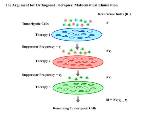
In summary, there are many avenues through which to approach future cancer therapies. We hope these new hallmarks depicting the stress phenotypes of cancer cells will both improve our understanding of the efficacy of existing therapies and provide rationales for new therapeutics. Cancer therapeutic targets can emerge through both the analysis of classical oncogenes and the analysis of a new class of genes that constitute NOA. New genome-wide efforts to approach cancer from a systems biology perspective hold great promise for identifying new targets for anti-cancer therapies. Finally, we propose that through the proper combination of orthogonal cancer therapies, it is possible to convert cancer from a death sentence into a manageable or even curable disease.


